Sipariş listesinde ürün yok
Ürün başarıyla alışveriş sepetinize eklendi
Sepetinizde 0 ürün bulunmaktadır. Sepetinizde 1 ürün bulunmaktadır.
Radyoloji
- Tıp Kitapları
- Acil Tıp
- Adli Tıp ve kriminoloji
- Aile Hekimliği
- Alerji ve İmmünoloji
- Anatomi Kitapları
- Anesteziyoloji ve Ağrı Kitapları
- Biyoloji ve Genetik Kitapları
- Biyomedikal Mühendisliği
- Biyokimya Kitapları
- Çocuk Cerrahisi
- Çocuk Sağlığı ve Hastalıkları Kitabı
- Çocuk ve Ergen Psikiyatrisi
- Dahiliye Kitapları
- Dermatoloji Kitapları
- Endokrinoloji Kitapları
- Farmakoloji Kitapları
- Fiziksel Tıp ve Rehabilitasyon
- Fizyoterapi, Rehabilitasyon ve Spor Hekimliği
- Fizyoloji Kitapları
- Gastroenteroloji Kitapları
- Geleneksel ve Tamamlayıcı Tıp
- Genel Cerrahi Kitapları
- Geriatri
- Göz Hastalıkları
- Göğüs Hastalıkları
- Göğüs Cerrahisi
- Halk Sağlığı Kitapları
- Hematoloji Kitapları
- Histoloji ve Embriyoloji Kitapları
- İnfeksiyon Hastalıkları
- Kadın Hastalıkları ve Doğum
- Kardiyoloji Kitapları
- Kalp Damar Cerrahisi
- Kulak Burun Boğaz Hastalıkları
- Mikrobiyoloji immunoloji Kİtapları
- Nöroşirürji
- Nefroloji
- Nöroloji
- Nükleer Tıp
- Onkoloji
- Ortopedi ve Travmatoloji
- Patoloji Kitapları
- Plastik Cerrahi
- Psikiyatri
- Radyasyon Onkoloji
- Radyoloji
- Romatoloji
- Sağlıklı Yaşıyoruz
- Spor Hekimliği
- Tıp Tarihi ve Tıp Etiği
- Tıbbı İstatistik Araştırma
- Tıp ve Sağlık Hukuku
- Tıbbi Laboratuvar Deney Bilimi
- USMLE & Board Review
- Uyku Tıbbı
- Üroloji
- Yoğun Bakım
- Diş Hekimliği Kitapları
- Eczacılık Kitapları
- Beslenme ve Diyet Kitapları
- Veteriner Hekimlik
- DUS Kitapları
- DUS Akademi Konu Kitapları Serisi
- DUS için Açıklamalı Deneme Sınavları Serisi
- DUS Spot Bilgiler Serisi
- Miadent Konu Kitapları Serisi
- Prodent Soru Kitapları Serisi
- DUS Çıkmış Soru Kitapları
- DUSDATA Şampiyonların Notu
- DUS Review Serisi
- DUSDATAMAX Soru Kitapları Serisi
- DUS Akademi Soru Kitapları Serisi
- Diğer Kitapları Serisi
- TUS Kitapları
- Çıkmış TUS Soru Kitapları
- 41 Deneme Serisi
- MEDOTOMY Serisi
- Tusmer
- Klinisyen Konu Kitapları Serisi
- Optimum Serisi
- Premium Serisi
- PRETUS Deneme Sınavları Serisi
- ProspekTUS Serisi
- Klinisyen Soru Kitapları Serisi
- Tusdata Ders Notları
- Tıbbi İngilizce
- Vaka Soruları Serisi
- Tüm Tus Soruları
- Hızlı Tekrar Serisi
- UTS Serisi
- KAMP ÖZEL NOTLARI
- Meditus Serisi
- YDUS Kitapları
- Hemşirelik ve Ebelik kitapları
- HEMŞİRELİK / halk sağlığı
- HEMŞİRELİK / Hemşirelik Esasları
- HEMŞİRELİK / İç Hastalıkları
- HEMŞİRELİK / Cerrahi Hastalıkları
- HEMŞİRELİK / Kadın hastalıkları ve doğum Ebelik
- HEMŞİRELİK / Ruh Sağlığı ve Hastalıkları
- HEMŞİRELİK / Hemşirelikte Eğitim
- HEMŞİRELİK / Çocuk Sağlığı ve Hastalıkları
- HEMŞİRELİK / Acil tıp hemşireliği
- SAĞLIK BİLİMLERİ
- Çocuk Gelişimi
- Sağlık Yöneticiliği
- Optisyenlik
- Odyoloji
- Saç Bakımı ve Güzellik Hizmetleri
- Anestezi Teknikerliği
- Tıbbi Dökümantasyon ve Sekreterlik
- Tıbbi Laboratuvar Teknisyenliği
- İş Sağlığı ve Güvenliği
- Ergoterapi
- Ağız ve Diş Sağlığı Teknisyenliği
- Dil ve Konuşma Terapisi
- İlk ve Acil Yardım Teknikeri (Paramedik)
- Radyoloji Teknisyenliği
- EĞİTİM BİLİMLERİ
- Değerler Eğitimi
- Eğitim Programları ve Öğretim
- Eğitim Psikolojisi
- Eğitim Yönetimi ve Denetimi
- Eğitimde Drama
- Eğitim Temelleri
- Eğitim Teknolojileri
- Okul Öncesi Eğitim
- Ortaokul Öğretmenliği
- Öğretmenlik Eğitimi Bölümleri
- Ölçme ve Değerlendirme
- Özel Eğitim
- Psikolojik Danışmanlık ve Rehberlik
- Sınıf Öğretmenliği
- Sınıf Yönetimi Etkili Öğretim
- Yabancı Dil Eğitimi
- İLETİŞİM
- İŞLETME
- İKTİSAT / EKONOMİ / MALİYE
- MİMARLIK - SANAT
- BİLİM TEKNİK
- MÜHENDİSLİK - TEKNİK
- FEN BİLİMLERİ
- ÇOCUK VE GENÇLİK KİTAPLARI
- BEŞERİ/SOSYAL BİLİMLER
- ÇEVRE ve YER BİLİMLERİ
- GIDA TARIM ve HAYVANCILIK
- BİYOMEDİKAL MÜHENDİSLİĞİ
- SEYAHAT TURİZM
- SOSYAL ÇALIŞMALAR
- SPOR BİLİMLERİ
- YÖNETİM - SİYASET - ULUSLARARASI İLİŞKİLER
- SINAVLAR HAZIRLIK
- ÖNERİLEN ÜRÜNLER
- Çok Satan Romanlar
- E-Kitaplar
- AYBAK
- Kırtasiye
- Yabancı Dil Eğitimi
- AYBAK 2025 Bahar
 Daha büyük görüntüle
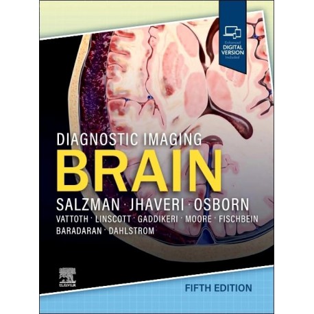
 Diagnostic Imaging Brain, 5th Edition
9780443380198
BU KİTAP İÇİN ÖN SİPARİŞ ALINMAKTADIR. TESLİM SÜRESİ 6 - 8 HAFTADIR. BİLGİ ALMAK İÇİN MAĞAZAMIZI ARAYINIZ
1 Adet
19 642,08 TL
15 713,66 TL
-20%
KDV Hariç: 15 713,66 TL
Dikkat: Stoktaki son ürün
- Yorum Yaz
Covering the entire spectrum of this fast-changing field, Diagnostic Imaging: Brain, fifth edition, is an invaluable resource for neuroradiologists, general radiologists, and trainees―anyone who requires an easily accessible, highly visual reference on today’s brain imaging. Drs. Miral Jhaveri, Karen L. Salzman, and a diverse group of neuroimaging experts provide updated information on more than 300 brain and CNS conditions to help you make informed decisions at the point of care. The text is lavishly illustrated, delineated, and referenced, making it a useful learning tool as well as a handy reference for daily practice.
- Provides authoritative, comprehensive guidance on both pathology-based and anatomy-based diagnoses to help you diagnose the full range of brain and CNS conditions
- Contains numerous new diagnoses and new chapters on such topics as inflammatory and demyelinating diseases, metabolic conditions, and CNS tumors
- Reflects significantly revised brain tumor categories based on the WHO CNS Classification and the Consortium to Inform Molecular and Practical Approaches to CNS Tumor Taxonomy, including major changes that advance the role of molecular diagnostics in CNS tumor classification
- Discusses key topics such as advances in treatment of Alzheimer’s disease with the approval of monoclonal antibody (MAB) immunotherapy, CNS manifestations of SARS-CoV-2, and recent advances in vessel wall imaging
- Features nearly 3,100+ high-quality print images (with an additional 4,700+ images in the complimentary eBook), including radiology images, full-color medical illustrations, clinical images, gross pathology photographs, and histology images
- Increases your ability to communicate imaging findings effectively with other neuroscientists, including oncologists and neurosurgeons
- Uses succinct bulleted text and highly templated chapters for quick comprehension of essential information at the point of care
- Includes an eBook that allows you access to everything in the print version as well as additional images, text, and references, with the ability to search, customize your content, make notes and highlights, and have content read aloud
SECTION 1: CONGENITAL MALFORMATIONS
Approach to Brain Malformations
Chiari Malformations
Chiari 1
Chiari 2
Chiari 3
Hindbrain Malformations
Dandy-Walker Continuum
Rhombencephalosynapsis
Unclassified Cerebellar Dysplasias
Molar Tooth Malformations (Joubert)
Cerebellar Hypoplasia
Disorders of Diverticulation/Cleavage
Holoprosencephaly
Syntelencephaly (Middle Interhemispheric Variant)
Septo-Optic Dysplasia
Commissural Abnormalities
Malformations Of Cortical Development
Congenital Microcephaly
Congenital Muscular Dystrophy
Heterotopic Gray Matter
Polymicrogyria
Focal Cortical Dysplasia
Lissencephaly
Schizencephaly
Hemimegalencephaly
Familial Tumor/Neurocutaneous Syndromes
Neurofibromatosis Type 1
Neurofibromatosis Type 2
von Hippel-Lindau Syndrome
Tuberous Sclerosis Complex
Sturge-Weber Syndrome
Meningioangiomatosis
Basal Cell Nevus Syndrome
Hereditary Hemorrhagic Telangiectasia
Neurocutaneous Melanosis
Encephalocraniocutaneous Lipomatosis
Aicardi Syndrome
Li-Fraumeni Syndrome
Schwannomatosis
Turcot Syndrome
Ataxia-Telangiectasia
PHACES Syndrome
SECTION 2: TRAUMA
Introduction to CNS Imaging, Trauma
Primary Effects Of Cns Trauma
Scalp and Skull Injuries
Missile and Penetrating Injury
Epidural Hematoma, Classic
Epidural Hematoma, Variant
Acute Subdural Hematoma
Subacute Subdural Hematoma
Chronic Subdural Hematoma
Traumatic Subarachnoid Hemorrhage
Cerebral Contusion
Diffuse Axonal Injury
Subcortical Injury
Pneumocephalus
Abusive Head Trauma
Secondary Effects of CNS Trauma
Intracranial Herniation Syndromes
Posttraumatic Brain Swelling
Traumatic Cerebral Ischemia/Infarction
Brain Death/Death by Neurologic Criteria (BD/DNC)
Second-Impact Syndrome
Blunt Cerebrovascular Injury
Traumatic Carotid Cavernous Fistula
Chronic Traumatic Encephalopathy
Leptomeningeal Cyst (Growing Fracture)
SECTION 3: SUBARACHNOID
Hemorrhage and Aneurysms
Subarachnoid Hemorrhage and Aneurysms Overview
Subarachnoid Hemorrhage
Aneurysmal Subarachnoid Hemorrhage
Perimesencephalic Nonaneurysmal Subarachnoid Hemorrhage
Convexal Subarachnoid Hemorrhage
Superficial Siderosis, Classic
Superficial Siderosis, Cortical
Aneurysms
Saccular Aneurysm
Pseudoaneurysm
Vertebrobasilar Dolichoectasia
ASVD Fusiform Aneurysm
Non-ASVD Fusiform Aneurysm
Blood Blister-Like Aneurysm
SECTION 4: STROKE
Stroke Overview
Nontraumatic Intracranial Hemorrhage
Evolution of Intracranial Hemorrhage
Spontaneous Nontraumatic Intracranial Hemorrhage
Hypertensive Intracranial Hemorrhage
Remote Cerebellar Hemorrhage
Germinal Matrix Hemorrhage
Critical Illness-Associated Microbleeds
Atherosclerosis and Carotid Stenosis
Intracranial Atherosclerosis
Extracranial Atherosclerosis
Arteriolosclerosis
Nonatheromatous Vasculopathy
Aberrant Internal Carotid Artery
Persistent Carotid Basilar Anastomoses
Sickle Cell Disease
Moyamoya
Primary Arteritis of CNS
Miscellaneous Vasculitis
Reversible Cerebral Vasoconstriction Syndrome
Vasospasm
Systemic Lupus Erythematosus
Cerebral Amyloid Disease
Cerebral Amyloid Disease, Inflammatory
Amyloid Related Imaging Abnormalities (ARIA)
CADASIL
Behçet Disease
Susac Syndrome
Fibromuscular Dysplasia
Cerebral Ischemia and Infarction
Hydranencephaly
White Matter Injury of Prematurity
Neonatal Hypoxic-Ischemic Injury
Adult Hypoxic-Ischemic Injury
Hypotensive Cerebral Infarction
Childhood Stroke
Cerebral Hemiatrophy
Acute Cerebral Ischemia/Infarction
Subacute Cerebral Infarction
Chronic Cerebral Infarction
Multiple Embolic Cerebral Infarctions
Fat Emboli Cerebral Infarction
Cerebral Embolism, Air
Lacunar Infarction
Cerebral Hyperperfusion Syndrome
Dural Sinus Thrombosis
Cortical Venous Thrombosis
404 Deep Cerebral Venous Thrombosis
Dural Sinus and Aberrant Arachnoid Granulations
SECTION 5: VASCULAR MALFORMATIONS
Vascular Malformations Overview
CVMS With AV Shunting
Arteriovenous Malformation
Dural AV Fistula
Pial AV Fistula
Vein of Galen Aneurysmal Malformation
Cerebral Proliferative Angiopathy
CVMS Without AV Shunting
Venous Anomaly
Sinus Pericranii
Cavernous Malformation
Capillary Telangiectasia
SECTION 6: NEOPLASMS
Neoplasms Overview
Gliomas, Glioneuronal Tumors, and Neuronal Tumors
Adult-Type Diffuse Gliomas
Astrocytoma, IDH-Mutant
Oligodendroglioma, IDH-Mutant and 1p/19q-Codeleted
Glioblastoma, IDH-Wildtype
Pediatric-Type Diffuse Low-Grade Gliomas
Diffuse Astrocytoma, MYB- or MYBL1-Altered
Angiocentric Glioma
Polymorphous Low-Grade Neuroepithelial Tumor of Young
Diffuse Low-Grade Glioma, MAPK Pathway-Altered
Pediatric-Type Diffuse High-Grade Gliomas
Diffuse Midline Glioma, H3 K27-Altered
Diffuse Hemispheric Glioma, H3 G34-Mutant
Diffuse Pediatric-Type High-Grade Glioma, H3-
Wildtype and IDH-Wildtype
Infant-Type Hemispheric Glioma
Circumscribed Astrocytic Gliomas
Pilocytic Astrocytoma
High-Grade Astrocytoma With Piloid Features
Pleomorphic Xanthoastrocytoma
Subependymal Giant Cell Astrocytoma
Astroblastoma, MN1-Altered
Glioneuronal and Neuronal Tumors
Ganglioglioma
Gangliocytoma
Desmoplastic Infantile Ganglioglioma/Desmoplastic Infantile Astrocytoma
DNET
Diffuse Glioneuronal Tumor With
Oligodendroglioma-Like Features and Nuclear Clusters
Papillary Glioneuronal Tumor
Rosette-Forming Glioneuronal Tumor
Myxoid Glioneuronal Tumor
Diffuse Leptomeningeal Glioneuronal Tumor
Multinodular and Vacuolating Neuronal Tumor
Dysplastic Cerebellar Ganglioglioma (Lhermitte-Duclos Disease)
Central Neurocytoma
Extraventricular Neurocytoma
Cerebellar Liponeurocytoma
| ISBN | 9780443380198 |
| Basım Yılı | 2026 |
| Basım Sayısı | 5 |
| Sayfa Sayısı | 1340 |
| Yazar(lar) | Karen L. Salzman, MD, FACR and Miral D. Jhaveri, MD, MBA |


